Medical science has advanced dramatically over the past century, and one of its most powerful innovations is diagnostic imaging—often shortened to diag image. From simple X-rays to advanced MRI scans, diagnostic imaging allows doctors to look inside the human body without surgery, providing vital insights for accurate diagnosis and effective treatment.
This article explores what diag image means, the different types of diagnostic imaging, their applications, benefits, risks, and the future of imaging in healthcare.
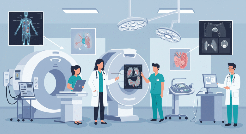
What Is a Diag Image?
A diag image is any medical image produced through diagnostic imaging techniques. These images help doctors:
-
Identify diseases and injuries.
-
Monitor treatment progress.
-
Guide medical procedures.
Without diag images, many conditions would remain hidden until too late. Today, diagnostic imaging is considered a cornerstone of modern medicine.
History of Diagnostic Imaging
The history of diag images dates back to 1895, when Wilhelm Conrad Roentgen discovered X-rays. This breakthrough revolutionized medicine by allowing physicians to see bones and foreign objects inside the body for the first time.
Since then, diagnostic imaging has evolved to include:
-
CT scans (1970s) – providing cross-sectional body images.
-
MRI scans (1980s) – using magnets and radio waves for detailed soft tissue imaging.
-
Ultrasound – using sound waves for safe, real-time imaging.
-
PET scans – offering metabolic and functional imaging.
Each advancement has expanded the ability of doctors to see more clearly and treat more effectively.
Types of Diag Images
1. X-rays
-
How it works: Uses electromagnetic radiation to capture images of bones and dense structures.
-
Common uses: Bone fractures, dental issues, chest conditions.
-
Advantages: Quick, inexpensive, widely available.
2. Computed Tomography (CT)
-
How it works: Combines X-ray images taken from multiple angles to create cross-sectional views.
-
Common uses: Detecting tumors, internal bleeding, organ injuries.
-
Advantages: High detail of bones and organs.
3. Magnetic Resonance Imaging (MRI)
-
How it works: Uses magnetic fields and radio waves to create detailed images of soft tissues.
-
Common uses: Brain scans, spinal issues, joint injuries.
-
Advantages: No radiation, excellent detail of soft tissues.
4. Ultrasound
-
How it works: Sound waves create live images of organs and tissues.
-
Common uses: Pregnancy monitoring, heart conditions, abdominal issues.
-
Advantages: Safe, portable, real-time imaging.
5. Positron Emission Tomography (PET)
-
How it works: Uses radioactive tracers to show metabolic activity.
-
Common uses: Cancer detection, brain disorders, heart disease.
-
Advantages: Shows function, not just structure.
6. Mammography
-
How it works: Specialized X-ray for breast tissue.
-
Common uses: Early detection of breast cancer.
-
Advantages: Proven tool in cancer screening programs.
Applications of Diag Images in Medicine
Diagnosis
Diag images help detect conditions such as:
-
Broken bones
-
Infections
-
Tumors and cancers
-
Blood clots
-
Neurological disorders
Treatment Planning
Before surgery or therapy, doctors use diag images to:
-
Map surgical areas.
-
Plan radiation treatments.
-
Guide minimally invasive procedures.
Monitoring Progress
Imaging helps track how well a treatment is working. For example:
-
Tumor shrinkage after chemotherapy.
-
Bone healing after a fracture.
Preventive Care
Regular imaging, like mammograms or low-dose CT scans, can catch diseases early before symptoms appear.
Benefits of Diag Images
-
Non-Invasive – No need for exploratory surgery.
-
Accurate Diagnosis – Increases precision in detecting conditions.
-
Faster Treatment – Quicker detection means earlier interventions.
-
Safer Procedures – Imaging guides surgeons in real-time.
-
Improved Outcomes – Better monitoring leads to more effective treatments.
Risks and Limitations of Diagnostic Imaging
While powerful, diag images come with certain risks:
-
Radiation Exposure – X-rays and CT scans expose patients to radiation (though doses are controlled).
-
Cost – Advanced scans like MRI and PET can be expensive.
-
Accessibility – Not always available in low-resource regions.
-
False Positives/Negatives – Imaging isn’t perfect; misinterpretation can occur.
-
Claustrophobia/Discomfort – Some patients struggle with MRI machines.
Doctors carefully weigh these risks against benefits before recommending scans.
Technological Advancements in Diag Images
The future of diagnostic imaging is being transformed by:
Artificial Intelligence (AI)
-
AI systems help detect abnormalities faster.
-
Algorithms can flag potential cancers or strokes with high accuracy.
3D Imaging
-
CT and MRI scans now produce 3D images, improving surgical planning.
Portable Imaging Devices
-
Handheld ultrasound devices make imaging accessible in remote areas.
Molecular Imaging
-
Allows visualization of cellular activity, offering earlier disease detection.
Ethical and Privacy Considerations
As diag images become digital, concerns about data privacy grow. Storing, sharing, and analyzing medical images must follow strict HIPAA and GDPR regulations to protect patient confidentiality.
Additionally, ethical questions arise about:
-
Over-imaging (unnecessary scans exposing patients to risk).
-
AI decision-making in diagnosis.
Healthcare providers must balance innovation with responsibility and ethics.
The Future of Diag Images
Looking forward, diagnostic imaging will continue to evolve with:
-
AI-powered diagnostics becoming routine.
-
Hybrid imaging (PET/CT, PET/MRI) offering structural + functional detail.
-
Tele-radiology expanding global access to expert analysis.
-
Preventive imaging programs helping reduce disease burden worldwide.
Ultimately, diag images will be central to personalized medicine, tailoring treatments to individual patients.
Conclusion
Diag images, or diagnostic imaging, are one of the most powerful tools in modern healthcare. From X-rays to advanced MRIs and PET scans, they allow doctors to see inside the body without invasive procedures, enabling accurate diagnoses, effective treatments, and better patient outcomes.
While challenges such as radiation exposure, costs, and accessibility remain, continuous innovation promises a future where diagnostic imaging is safer, smarter, and more widely available.
For patients and doctors alike, diag images represent a bridge between visible symptoms and hidden conditions—bringing clarity where it matters most.